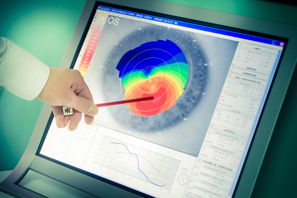
In the realm of optometry, technology has always played a pivotal role, and corneal topography is no exception. As a non-invasive medical imaging technique for mapping the surface curvature of the cornea, corneal topography has revolutionized the way eye health professionals diagnose, monitor, and treat a variety of ocular conditions.
What is Corneal Topography?
Corneal topography is a process that generates a detailed, three-dimensional map of the cornea's surface. The cornea is responsible for refracting most of the light coming into the eye. Therefore, any irregularities in its shape and smoothness can affect vision.
Corneal topography is an invaluable tool for detecting, diagnosing, and monitoring numerous corneal conditions, deformities, and irregularities. It is also instrumental in planning refractive surgery and evaluating its success. The data gathered from corneal topography can provide an extensive understanding of the cornea’s health and function, which is vital for many eye-related diagnoses and treatments.
How Does Corneal Topography Work?
Corneal topography works by measuring the light reflected off the cornea. It utilizes a computerized system that shines a series of concentric rings of light onto the cornea. The shape the light rings take when reflected back is captured and analyzed by a computer, creating a detailed map of the corneal surface.
The resulting map, often color-coded for easier interpretation, reveals minute details about the cornea's shape and curvature. Red areas on the map typically indicate steeper, or higher areas, while blue areas indicate flatter, or lower areas. This detailed information provides a comprehensive understanding of the cornea’s condition, enabling eye health professionals to make accurate diagnoses and treatment plans.
Common Uses for Corneal Topography
Corneal topography is used in a variety of contexts within eye care. It is commonly used to diagnose and manage a range of corneal conditions, such as keratoconus, a condition where the cornea thins and bulges into a cone-like shape. By creating a detailed map of the cornea, corneal topography can detect subtle changes in the cornea's surface, providing early diagnosis of conditions like keratoconus.
Additionally, corneal topography is indispensable in contact lens fitting, especially for patients with irregular corneas. The detailed map allows for a more accurate and comfortable fit, significantly improving the patient’s overall experience.
Benefits of Corneal Topography
The benefits of corneal topography are manifold. First, it provides exceptionally detailed and accurate information about the cornea's shape and condition. This allows for early detection of corneal irregularities, leading to timely interventions and improved patient outcomes.
Additionally, in the context of contact lens fitting, corneal topography is invaluable. It helps practitioners fit lenses with a high degree of accuracy, ensuring comfort and clear vision for the patient. This is particularly beneficial for patients with irregular corneas, who may otherwise struggle to find a comfortable and effective fit with traditional fitting methods.
Conclusion
Corneal topography is a powerful tool in the field of optometry and ophthalmology. By providing a comprehensive map of the cornea, it enhances the diagnosis, monitoring, and treatment of a variety of eye conditions. Whether detecting early signs of keratoconus, or fitting a contact lens, corneal topography proves its worth. It's clear that the benefits of this technology extend not only to eye health professionals but also, and most importantly, to patients, who ultimately enjoy improved eye health and vision outcomes.
To learn more on the benefits of corneal topography, contact Dau Family Eye Care at our office in St. John’s, Florida. Please call 904-713-2020 to schedule an appointment today.


